Sunday, August 29, 2010

Structures and Colours - Dermatoscopy Made Easy

When you look at a lesion you are seeking out  colours and structures. 

Colours in the skin are due to melanin pigment, blood, keratin and collagen and pigments added from the environment. Melanin can be in nevus cells or melanocytes and keratinocytes in the epidermis and also in macrophages and other tumour proliferating cells in the dermis. Melanin is black if very superficial in the epidermis, brown at the dermo epidermal junction , grey in macrophages in the papillary dermis and blue in the deeper nevus cells of a blue nevus or a melanoma metastasis or a pigmented basal cell carcinoma.

 Hemangiomas are red or purple  while Seb ks are brown, yellow or black if they have a lot of melanin in them. 

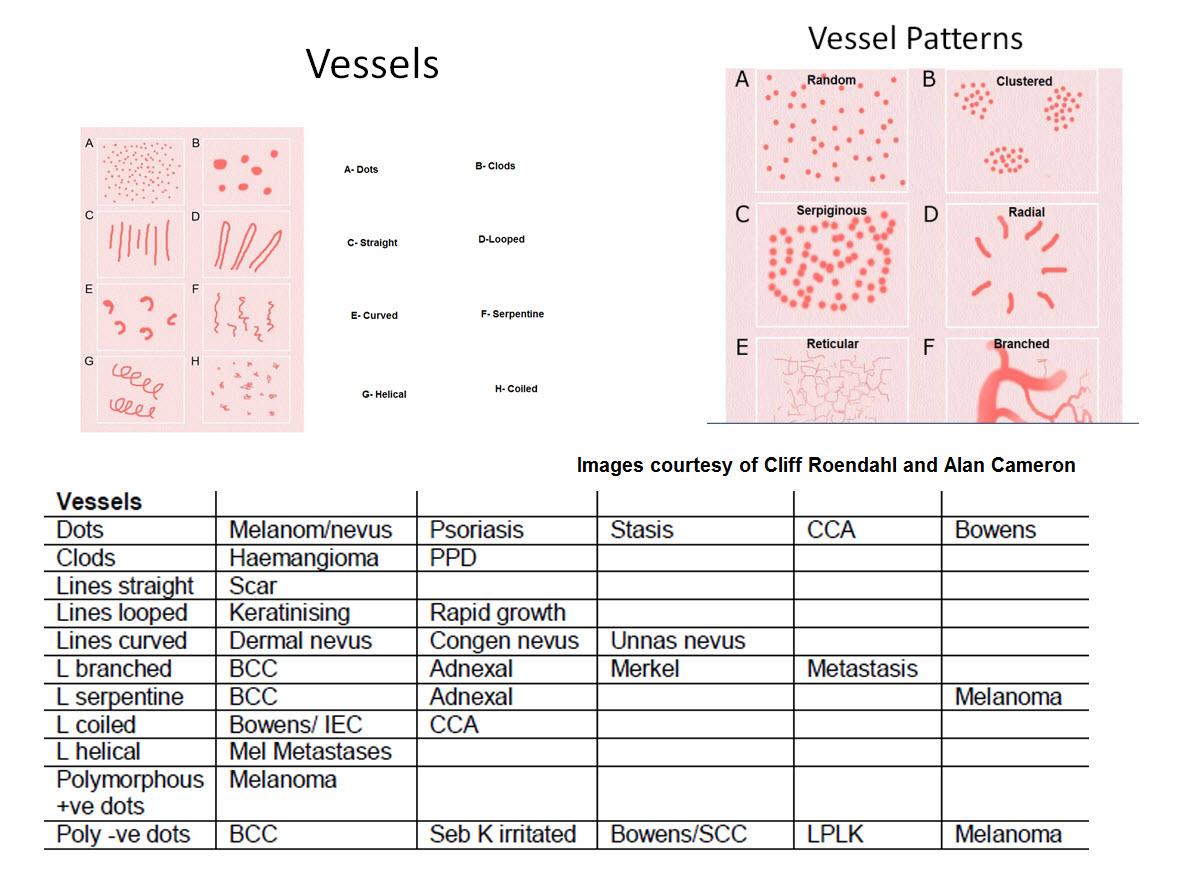
What about structures? Look for lines, circles, dots, clods or one colour structureless areas. With pink lesions you look at the vessels and background white lines to try to make a diagnosis .

Just look at the links opposite for each of these elements and what they represent.  Start with Lines and work your way down. If a particular diagnosis is mentioned that you are unfamiliar with then scroll down the links opposite and look it up. This way you will learn what each of the colours and structures might represent and that should help you to make the correct diagnosis. However remember the real question here is not the correct diagnosis but instead do I leave this lesion, cut it out or monitor it over say 3 months. Let me quickly add you do not monitor something that is raised and rapidly growing. You cut it out.

Using a dermatoscope allows you to quickly determine if a lesion is benign. If a pigmented lesion is one colour and symmetrical then ignore it. Move on to the next lesion. I am assuming you can recognise a typical  seborrhoeic keratosis and hemangioma. If not look first at the Seb k and hemangioma links opposite and learn the patterns. If it is pink look at the vessels.

Look at the table below to see which lesions commonly have which structures and the patterns in which these structures are arranged. Keep in mind that a symmetrical arrangement of structures is usually a benign lesion. You should also learn all the clues to melanoma. I have added them below but look at the link opposite to see good clinical examples. 

Take particular care of lesions that the patient says are new or have changed. Look at them particularly carefully. Know your patient's past history. If they already have had two melanomas then you excise any new pigmented lesion that is not obviously benign.

Take care with new rapidly growing pink papules. They usually all end up needing excised. See Amelanotic Melanoma

So pick up your dermatoscope and let us get started


Introduction.

Many of the ideas in this teaching blog come from the work of Harald Kittler, Scott Menzies, Cliff Rosendahl and Alan Cameron and the BLINCK algorithm is the work of Dr Peter Bourne.

View this YouTube video on the website. Click on the arrow and when it starts go to the bottom of the screen and change the 360 resolution to 1080 and then click on the box with the arrows pointing out to enlarge to full screen. These videos are recorded in high definition which makes for great viewing if you have a fast broadband connection. Press ESC on your keyboard to return to normal size.


If you want to view examples of these clues then click on the post opposite or click here


Which dermatoscope should I buy? and Using my Dermatoscope

Since the above was written 10 years ago Heine have updated the Delta 20 to have 
both polarised and non polarised light.  See the Heine website Heine also put out a Delta 30 but it was inadequate in comparison to the Delta 20S in showing up white lines in polarised mode so they are now putting out a Delta 30 Pro. If you can get your hands on a second hand Delta 20S you wont go wrong!

There is also now a DL5 dermatoscope from 3Gen. Magnetic rings allow easy coupling of small digital cameras to the dermatoscope to record images. See Australian DL5 distributer

You can also get attachments for smartphones from both Heine and 3Gen but I still prefer a dedicated setup.


Older but still excellent Heine Delta 20S

Heine Delta 30 Pro


DL 5 with optional ruler


Using your Dermatoscope

If someone has previously had a melanoma try to examine all pigmented lesions on their skin and any new pink lesions.

If the pigmented lesion is symmetrical and one colour move on. Look carefully at all dark pigmented lesions and any with varying pigmentation and all large pigmented lesions.

On the face with a pigmented lesion look for grey circles or partial grey circles as they often point to a lentigo maligna.

On the feet look at the edge of the pigmented lesion. If the pigment is in the furrows then the lesion is a benign acral nevus. Don’t concern yourself with the centre of the lesion which might be lattice or fibrillar. If however the edges show ridge pigmentation you have either an acral melanoma or subcorneal bleeding.

Look for polygons in large pigmented lesions on the trunk. They certainly should have you thinking of a lentiginous melanoma.

Pink raised lesions could be an amelanotic melanoma so look for polymorphous vessels but if ulceration consider BCC more likely with linear irregular vessels and sometimes white lines. White lines are also a feature of desmoplastic melanomas.  

Elevated pink growing lesions generally need to be excised irrespective of the dermatoscopy to exclude other tumours such as Merkel cell, Atypical Fibroxanthoma, Poorly differentiated SCC and skin metastases.

Pink flat lesions could be BCC (linear irregular branched vessels) , SCC in situ (dot or glomerular vessels), amelanotic melanoma (polymorphous and dot vessels , white lines)

The dermatoscope is best for diagnosing benign lesions. 

Hemangiomas have vascular globules or clods.

Seborrhoeic keratoses have orange, brown or white clods. There are a wide range of variants though and irritated light coloured se bks can look very like an SCC. Melanoacanthomas can be so dark you think they are melanomas. Clear cell acanthomas are pink with a distinctive vascular pattern.

Dermatofibromas have a white collagen centre with a surrounding reticular network looking like a pigmented lesion but there are other variants.

Adnexal tumours including warts are usually skin coloured and some are scaly. The dermatoscope can show the thrombosed capillaries of a wart but there are few specific dermatoscopic patterns for these tumours and histology is best to define them.

Some benign nevi are easier to diagnose dermatoscopically than others. The easy ones to diagnose are blue nevi, congenital nevi, Dermal nevi, Reed and Spitz nevi.  Clinically atypical nevi that are later diagnosed histologically dysplastic can have overlap features of a melanoma and are rightly excised.

The other common skin cancers are basal and squamous cell carcinomas. You don’t see a pigment network in a BCC  but pigmented BCCs will show dark blue clods of pigment and grey dots.

SCCs have lots of keratin especially when histologically well differentiated so look for looped vessels surrounded by keratin or white circles in hair follicles. The superficial in situ SCC or Bowen’s disease will show dot or glomerular vessels as its main dermatoscopic feature.

Lastly the dermatoscope can be used in medical dermatology in a wide range of diseases to assist in the clinical diagnosis.

Colours in Dermoscopy

View this YouTube video on Colours. Click on the arrow and when it starts go to the bottom of the screen and change the 360 resolution to 1080 and then click on the box with the arrows pointing out to enlarge to full screen. These videos are recorded in high definition. Press ESC on your keyboard to return to normal size.


Black combined with brown is usually due to melanin whereas black plus red is usually due to bleeding.


Yellow also is seen in Xanthomas and Xanthogranuloma 

\White can be seen in Mollusca, Pilomatricoma and Gouty tophi with the various deposits in the skin.


Kittlerian Terminology

View this YouTube video on Kittlerian terminology. Click on the arrow and when it starts go to the bottom of the screen and change the 360 resolution to 1080 and then click on the box with the arrows pointing out to enlarge to full screen. These videos are recorded in high definition. Press ESC on your keyboard to return to normal size.


Lines


View these YouTube videos on the different types of lines. Click on the arrow and when it starts go to the bottom of the screen and change the 360 resolution to 1080 and then click on the box with the arrows pointing out to enlarge to full screen. These videos are recorded in high definition. Press ESC on your keyboard to return to normal size.


Lines take precedence over other structures and lines reticular is a common structure because we look at a lot of nevi. Remember it is made up of the arrangement of pigmented cells over the dermal papillae (clear holes) and along the sides of the rete ridges (pigmented network).


Lines radial peripheral represent confluent junctional nests of melanocytes that are rapidly proliferating, They are a feature of Spitz and Reed nevi but when asymmetrically distributed around a pigmented lesion then you should consider melanoma.
Pseudopods are lines radial peripheral with a knob joined to the streak at the end. They are also seen in Spitz, Reed nevi and melanoma. If asymmetrically distributed then consider melanoma and biopsy remove.


View this YouTube video.


View this YouTube video on Lines Parallel


White Lines

White lines are seen in mainly BCCs, invasive melanomas and in Dermatofibromas and scars and some Spitz nevi. They are best seen with a polarising dermatoscope. They may cross each other at right angles.  Some people believe these white lines are just polarising artefacts but I doubt it. Collagen in the dermis is birefringent to polarised light. Sometimes these white lines on a pink background are all you see in a desmoplastic melanoma!